Some basic knowledge about the normal human brain will assist to fully appreciate what is happening in the Alzheimer brain
THE HUMAN BRAIN:
- Weighs about 3 pounds
- Vast network of blood vessels
- billions of capillaries carry oxygen, glucose, nutrients and hormones to the brain
- Composed of two main classes of cells:
- neurons (about a billion) and glia (glial cells)
NEURONS: THE COMMUNICATORS
These cells have four morphological defined regions.
- Cell body
- Dendrites
- Axons
- Axon or presynaptic terminals
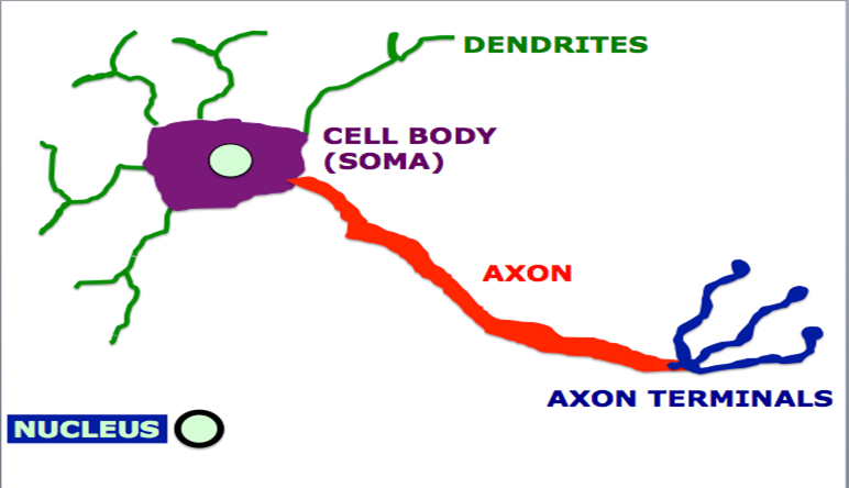
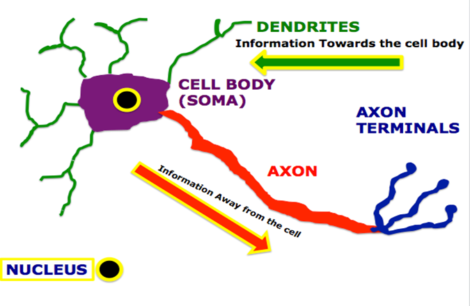
The neuron is the functional unit of the nervous system.
The cell body is the metabolic center of the neuron. The nucleus, which is located in the cell body most of the protein synthesis occurs in the cell body.
Dendrites branch out from the cell body and their role is to receive messages from axons and other neurons.
The axon extends out from the cell body. Depending on the part of the nervous system axons can be very long processes up to a meter long. The axon carries messages away from the neuron.
Axon or presynaptic terminals are specialized swellings at the end of fine branches of the axon. These terminals end in close proximity to dendrites of postsynaptic neurons, which receives messages from the presynaptic terminal.
Each neuron communicates with thousands of nerve cells through its axons and dendrites. Within the brain groups of neurons have specific roles. Some are involved with thinking, learning, emotions, and memory. Others receive information the eyes, ears and skin this is known as sensory information. Other groups of neurons control and stimulate muscle activity.
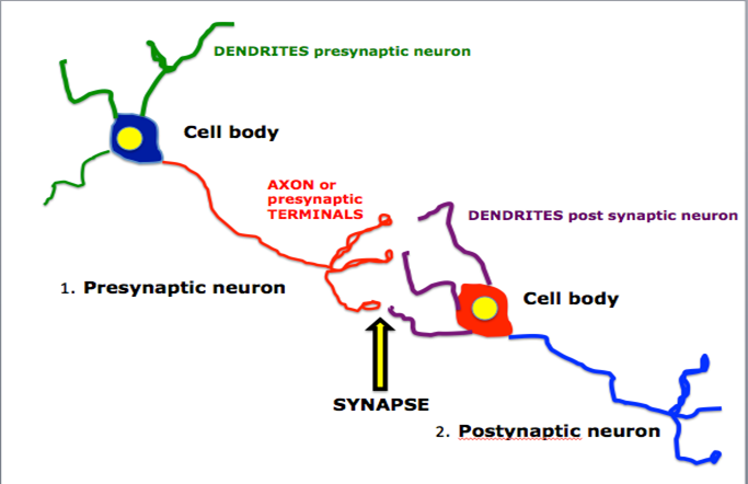
Neurons communicate with other neurons. Dendrites receive an incoming signal (electrical or chemical) which generates an nerve impulse or action potential in the cell body. These impulses then travel to the end of the axon, which trigger the release of chemical messengers or neurotransmitters. This information passes from the axon of the presynaptic neuron to the dendrites of the postsynaptic neuron. The site where the axon or presynaptic terminals end in proximity of the receiving dendrites is known as the synapse.
SYNAPSE
- Cell sending information out is the presynaptic neuron
- Cell receiving information is the postsynaptic neuron
- At the synapse there is no contact between the presynaptic neuron and postsynaptic neuron. This space is known as the synaptic cleft
- One neuron can form as many as 7000 synapses with other neurons.
- Neurons can form new synapses or reinforce synaptic connections in response to life experiences. Changes in neuronal connections are the basis of learning.
The synapse is where neurotransmitters are released from the axon terminal of the presynaptic neuron. Neurotransmitters are stored in vesicles. Each vesicle contains thousands of neurotransmitter molecules. The vesicles fuse with the membrane of the presynaptic neuron and release their content via exocytosis. Once released neurotransmitters bind to specific receptors on the postsynaptic neuron, which in turn elicits a response. Some neurotransmitters inhibit nerve cell function. This means that the receiving nerve cell will not send a signal down its axon. Other neurotransmitters excite or stimulate nerve cells meaning that the receiving neuron will send electrical signals down to axon to more receiving neurons in the pathway.
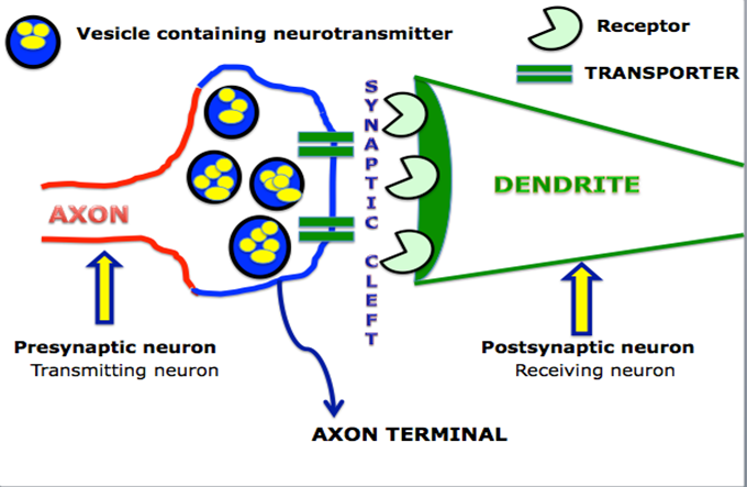
Neurotransmitters that did not bind to the receptor:
- Can be degraded by enzymes in the synapse or
- Taken back up into the presynaptic terminal by a transporter or reuptake pump
- Here it is either destroyed or repackaged into new vesicles.
Communication is one of the biological process which is vital for the function and survival of neurons. The other two are:
- METABOLISM
This refers to all chemical reactions taking place in a cell to survive and function. These reactions require glucose and oxygen, which is supplied by the blood circulating in the brain. The brain weighs 3 pounds and contains the richest blood supply of any organ and consumes 20 % of the energy used by the human body. - REPAIR, REMODELING & REGENERATION
Neurons are programmed to live at least 100 years in the human brain. To achieve this survival they must constantly maintain and repair themselves. Their synaptic connections are continuously remodeled based on the amount of stimulation they receive from other neurons. Neurons can strengthen, weaken or even break connections with groups of neurons to reestablish new connections. Remodeling of synaptic connections are considered to be important for learning, memory and even brain repair.
GLIA THE SUPPORT CELLS
There are four types of glial cells: microglia, oligodendrocytes, astrocytes and Schwann cells. Neurons survive and function with the help and support of glial cells, the other main type of cell in the brain. Glial cells hold neurons in place, provide them with nutrients, rid the brain of damaged cells and other cellular debris, and provide insulation to neurons in the brain and peripheral nervous system. To carry out these functions, glial cells often act in collaboration with blood vessel cells, which in the brain have specialized features not found in blood vessels elsewhere in the body. Together, glia and blood vessel cells regulate the delicate chemical balance within the brain to ensure optimal neuronal function.
KEY BRAIN REGIONS
There are many parts of the human brain each can be considered to have specific roles while communicating among each other.
1. CEREBRUM
The cerebrum is the seat of many of the higher mental functions, such as memory and learning, language, and conscious perception. The cerebrum is divided into right and left hemispheres that are connected by the corpus callosum.
In various ways, researchers have discovered that the two halves do have some specialization. It is the left hemisphere that relates to the right side of the body (generally), and the right hemisphere that relates to the left side of the body. Also, it is the left hemisphere that usually has language, and seems to be primarily responsible for similar systems such as math and logic. The right hemisphere has more to do with things like spatial orientation, face recognition, and body image. It also seems to govern our ability to appreciate art and music.
2. CEREBRAL CORTEX
The cerebral cortex is the thin layer of gray matter on the outside of the cerebrum. It is approximately 2-3 millimeter thick in most regions. It is highly and tightly folded to fit within the limited space of the cranial vault. These folds give the brain a very wrinkled appearance. The folding creates grooves known as sulci and bumps known as gyri. The folding increases the surface of the brain.
The cerebral cortex is the area where all information such as sensory, volunteer control over movement, cognitive function including thinking, learning, memory, and decision making is processed.
Billions of nerve cells make up the gray surface of the cerebrum. White nerve fibers underneath carry signals between the nerve cells and other parts of the brain and body.
Most of the cortex is known as the neocortex and consists of 6 layers. Older parts of the cortex are referred to as the allocortex and are located in the medial temporal lobe. These areas are concerned with olfaction (sense of smell),and emotions. The allocortex has 2 components : the paleocortex and the important archicortex which consists of the hippocampus. The hippocampus is an essential structure since it is involved with memory.
3. LOBES
As seen in the figures below, the cerebral hemispheres are further subdivided into 4 lobes: Frontal, Parietal, Temporal and Occipital. The lobes all have different functions.
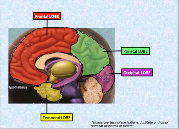
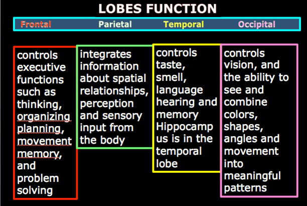
4. OTHER IMPORTANT BRAIN REGIONS CAN BE SEEN IN THE FIGURE BELOW
Cerebellum is located above the brainstem and beneath the occipital lobe. It plays a role in balance and coordination.
Brainstem is located at the base of the brain. It connects the spinal cord with the rest of the brain. Though it is smallest part of the brain it plays a critical role in our survival as it controls heart rate, blood pressure, and breathing. Sleep is also regulated in the brainstem. It also relays all information between the brain and spinal cord such the control of movement and sensory input.
Amygdala is located in the temporal lobe and is involved in processing and remembering strong emotions such as fear anxiety.
Hippocampus is buried in the temporal lobe and is extremely important for learning and memories.
Thalamus is located at the top of the brainstem and is responsible for processing sensory and limbic information before it is conveyed to the cortex.
Hypothalamus is located under the thalamus and controls activities such as temperature, and food intake.
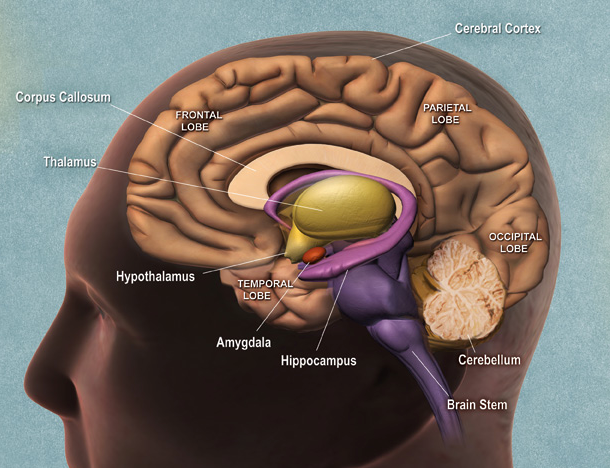
Figure shows other important brain regions Note the location of the hippocampus and amygdala in the medial temporal lobe.
“Image courtesy of the National Institute on Aging/National Institutes of Health”
Sources of information from the following.For more information please consult the following:
http://www.alz.org/braintour/signal_activity.asp
http://science.education.nih.gov/supplements/nih2/addiction/guide/guide_toc.htm
http://www.nia.nih.gov/alzheimers/publication/introduction/about-book
http://www.alzheimers.org.uk/braintour
http://www.alz.org/alzheimers_disease_4719.asp?type=alzFooter
